Draw A Microscope And Label
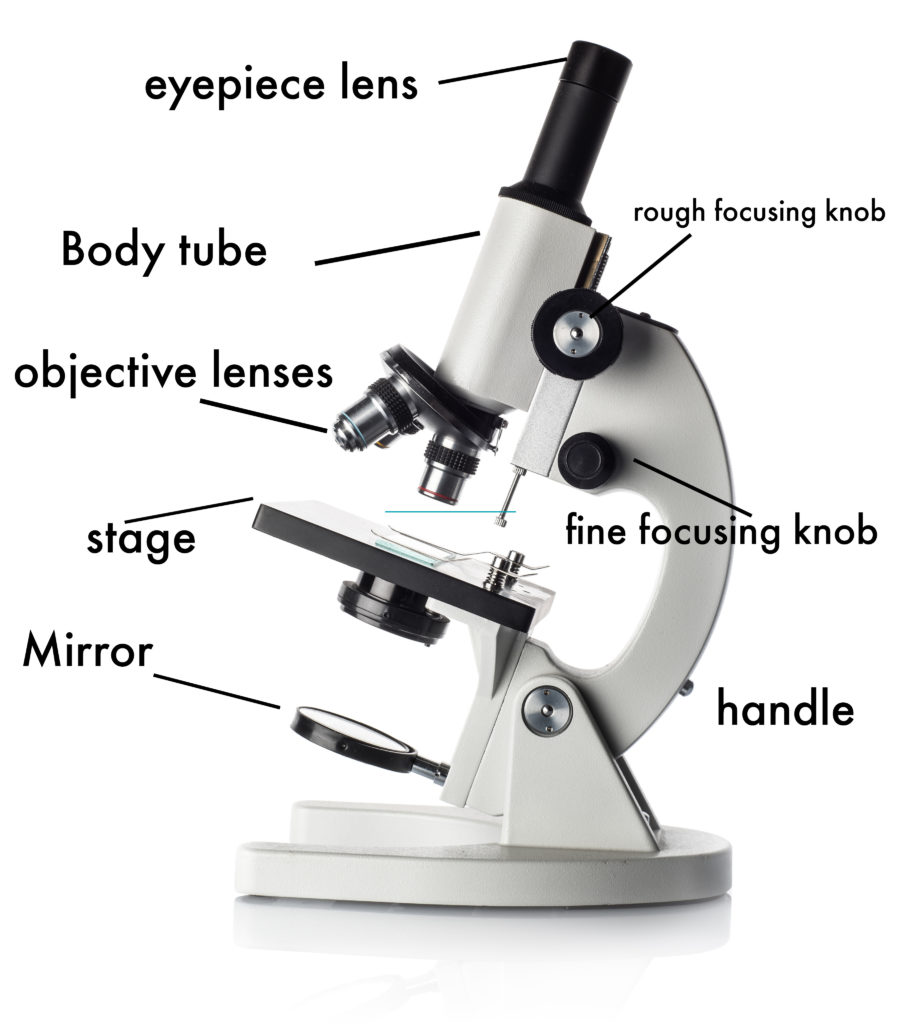
Draw A Microscope And Label - Web ready to take your drawing skills to the next level? The upper part of the microscope that houses the optical elements of the unit.; Label the parts of the microscope with answers (a4) pdf print version. Label the parts of the microscope (a4) pdf print version. Be sure to indicate the magnification used and specimen name. Using arrows and textables label each part of the microscope and describe its function. Web how to draw a microscope 🔬. Let’s tell you how to do it: Web use this interactive to identify and label the main parts of a microscope. There are three structural parts of the microscope i.e. Label the cell wall, cell membrane, cytoplasm, and chloroplasts in your lab manual. Web these labeled microscope diagrams and the functions of its various parts, attempt to simplify the microscope for you. Draw the objective lenses 1.5 step 5: There are six printables available. Web ready to take your drawing skills to the next level? This activity has been designed for use in homes and schools. Web today, we're learning how to draw a cool microscope!👩🎨 join our art hub membership! There are three structural parts of the microscope i.e. Label the cell wall, cell membrane, cytoplasm, and chloroplasts in your lab manual. There are six printables available. Diagram of parts of a microscope. Take a look at your microscope slide and start with the basic shapes and outlines of the objects you see. The base is attached to a frame (arm) that is connected to the head of the device.the base of the microscope provides stability to the device and allows the user’s. The rectangle should be. Shape the microscope head 1.3 step 3: It is also called a body tube or eyepiece tube. Web when labeling a microscope, accuracy is key. Take a look at your microscope slide and start with the basic shapes and outlines of the objects you see. This activity has been designed for use in homes and schools. It is also called a body tube or eyepiece tube. The microscope layout, including the blank and answered versions are available as pdf downloads. Use a landscape poster layout (large or small). Web today, we're learning how to draw a cool microscope!👩🎨 join our art hub membership! Download the diagrams and practice labeling the different parts of these. If you are using a graticule slide (a microscope slide with millimeter grid lines), lightly sketch a grid over your circle. This can be done with pencil and paper or with digital drawing tools. The base is attached to a frame (arm) that is connected to the head of the device.the base of the microscope provides stability to the device. Web labeling the parts of the microscope. Label the cell wall, cell membrane, cytoplasm, and chloroplasts in your lab manual. Web the individual parts of a compound microscope can vary heavily depending on the configuration & applications that the scope is being used for. It is also called a body tube or eyepiece tube. Web table of contents 1 how. Shape the microscope head 1.3 step 3: Be sure to indicate the magnification used and specimen name. It will take 9 steps in total to complete the drawing. Web how to draw a microscope 🔬. Web labeled diagram of a compound microscope major structural parts of a compound microscope optical components of a compound microscope eyepiece eyepiece tube objective lenses. Use a landscape poster layout (large or small). Web this exercise is created to be used in homes and schools. Web use this interactive to identify and label the main parts of a microscope. There are six printables available. Web labeled diagram of a compound microscope major structural parts of a compound microscope optical components of a compound microscope eyepiece. Outline the slide platform 1.6 step 6: Common compound microscope parts include: Using arrows and textables label each part of the microscope and describe its function. The part that is looked through at the top of the compound. Continue follow my channel and like, share,comment also. It is important to make sure you have the correct information before labeling your microscope. Web this exercise is created to be used in homes and schools. Use a landscape poster layout (large or small). In this interactive, you can label the different parts of a microscope. Web labeled diagram of a compound microscope major structural parts of a compound microscope optical components of a compound microscope eyepiece eyepiece tube objective lenses nosepiece specimen stage coarse and fine focus knobs rack stop illuminator condenser abbe condenser iris diaphragm condenser focus knob. Label the parts of the microscope (a4) pdf print version. There are three structural parts of the microscope i.e. And drop the text labels onto the microscope diagram. First and foremost, we have a labeled microscope diagram, available in both black and white and color. Shape the microscope head 1.3 step 3: Web the goal is to complete a drawing of a microscope by creating each part one part at a time. Search for a diagram of a microscope. Download the diagrams and practice labeling the different parts of these. Here are some steps to help ensure that you label your microscope correctly: The base is attached to a frame (arm) that is connected to the head of the device.the base of the microscope provides stability to the device and allows the user’s. Perfect for students or anyone.
Compound Microscope Parts Labeled Diagram and their Functions Rs

Microscope Drawing And Label at GetDrawings Free download

Parts of a Microscope Labeling Activity

Simple Microscope Drawing at GetDrawings Free download

Parts of a microscope with functions and labeled diagram

How to draw Microscope diagram for beginners step by step YouTube

Simple Microscope Drawing at GetDrawings Free download
How to Use a Microscope

Monday September 25 Parts of a Compound Light Microscope

Labeled Microscope Diagram Tim's Printables
Web Labeling The Parts Of The Microscope.
Web These Labeled Microscope Diagrams And The Functions Of Its Various Parts, Attempt To Simplify The Microscope For You.
The Upper Part Of The Microscope That Houses The Optical Elements Of The Unit.;
The Rectangle Should Be As Tall As You Want The Microscope To Be.
Related Post: